Key Details
What to know before your visit
A key screening tool for early detection of breast cancer.
Used for routine screening and diagnostic assessment of breast concerns.
Often paired with breast ultrasound when additional imaging is needed.
About the Exam
How Mammography supports diagnosis
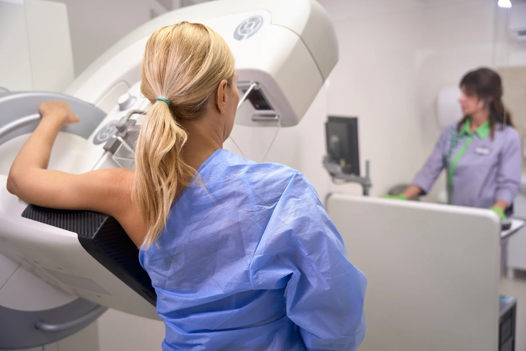
Mammography is a specialized medical imaging technique that uses low-dose X-rays to examine breast tissue. It is the most effective screening tool for the early detection of breast cancer, often identifying abnormalities before physical symptoms develop. There are two main types: screening mammography for routine checks in women without symptoms, and diagnostic mammography for evaluating specific breast concerns, such as lumps or pain. Mammography is safe, quick, and plays a critical role in saving lives through early diagnosis and treatment.
How to Prepare:
To prepare for your mammogram at Erin Diagnostic Imaging, please avoid using deodorant, antiperspirant, powders, lotions, or perfumes on your underarms and chest on the day of your appointment, as these can appear on the images and affect accuracy. You will be asked to undress from the waist up and wear a gown provided by the clinic.
We recommend wearing a two-piece outfit for comfort and convenience. If you have had previous mammograms at another clinic, please bring or arrange for those images to be sent for comparison, as this can help the radiologist with a more accurate assessment.
Bring your requisition, if needed and health card. Our team will guide you through the process and ensure your comfort throughout the exam.
What Happens During my Mammogram:
During your mammogram at Erin Diagnostic Imaging, you will be welcomed by our team and asked to change into a gown, removing clothing from the waist up. The technologist will ask you to confirm your requisition and health information and explain the procedure and position you in front of the mammography unit to ensure the best possible images are obtained.
Each breast is gently compressed between two plates for a few seconds while the X-ray image is taken. This compression is important as it helps spread out the breast tissue, reduces motion, and allows for clearer, more detailed images while using a low dose of radiation. You may feel some temporary pressure during this step, but it only lasts a few seconds per image.
Multiple images are taken from different angles to ensure a complete assessment. Once the exam is finished, you can resume your normal activities immediately. Your images will be reviewed by a radiologist, and the results will be sent to your referring healthcare provider.
Related Services
Explore Mammography services
Appointments
Ready to book Mammography?
Submit your requisition online or call the clinic and our team will help coordinate your visit.