Key Details
What to know before your visit
Also known as cardiac ultrasound.
Helps assess heart valves, function, structure and blood flow.
Little to no special preparation is usually required.
About the Exam
How Echocardiography supports diagnosis
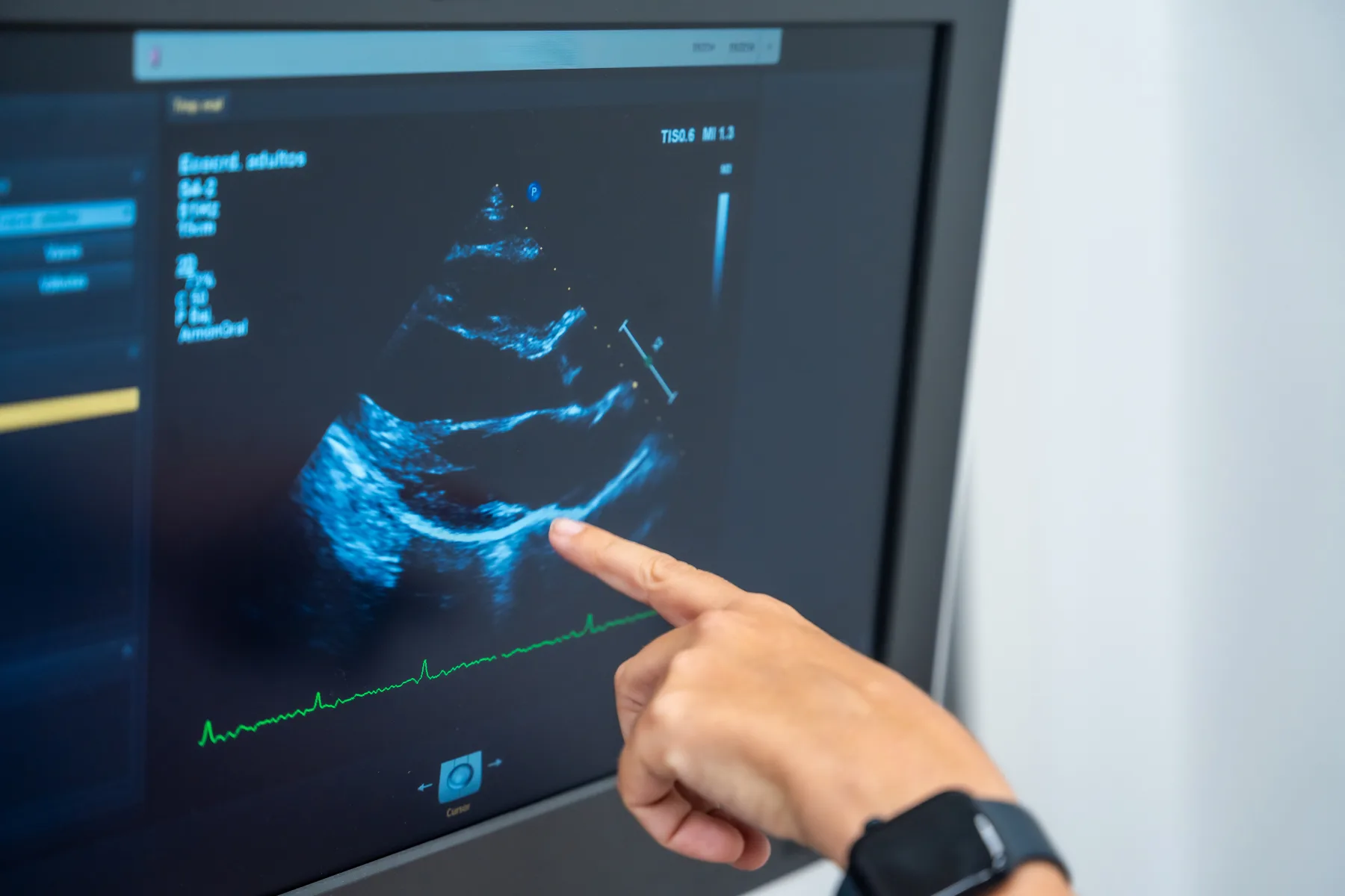
Erin Diagnostic Imaging is partnered with cardiologists within the community. Echocardiography, or cardiac ultrasound, is a non-invasive imaging technique that uses high-frequency sound waves (ultrasound) to produce real-time images of the heart. It allows physicians to assess the heart’s structure, function, and blood flow, making it essential for diagnosing conditions such as heart valve disorders, heart failure, congenital heart defects, and cardiomyopathy. Echocardiograms are safe, painless, and do not use ionizing radiation, making them a preferred method for routine cardiac evaluation.
How to Prepare:
Upon referral you will be booked with our partner cardiology clinic for a consultation and appointment for echocardiogram. To prepare for your echocardiogram, little to no special preparation is usually required. You may eat, drink, and take your regular medications unless your healthcare provider has given you different instructions.
We recommend wearing comfortable clothing, as you may be asked to change into a gown for the examination. Please avoid applying lotions or oils to your chest on the day of your appointment, as this can interfere with the ultrasound images.
Be sure to bring your requisition and health card to your appointment and arrive a few minutes early for check-in. If you have any questions about your exam preparation, our team will be happy to assist you.
What Happens During my Echocardiogram:
During your echocardiogram, you will be asked to lie comfortably on an examination table while a technologist performs an ultrasound of your heart. Small electrodes may be placed on your chest to monitor your heart rhythm during the exam.
A gel will be applied to your chest, and a handheld device called a transducer will be gently moved over the area to capture real-time images of your heart’s structure and function using sound waves. The technologist will obtain detailed images to assess how your heart chambers, valves, and blood flow are functioning.
The exam is painless, non-invasive, and typically takes between 30 to 60 minutes. Once the examination is complete, you may resume your normal activities. Your images will be reviewed by a physician, and the results will be sent to your referring healthcare provider.
Appointments
Ready to book Echocardiography?
Submit your requisition online or call the clinic and our team will help coordinate your visit.