Service Information
About Anatomy Ultrasound
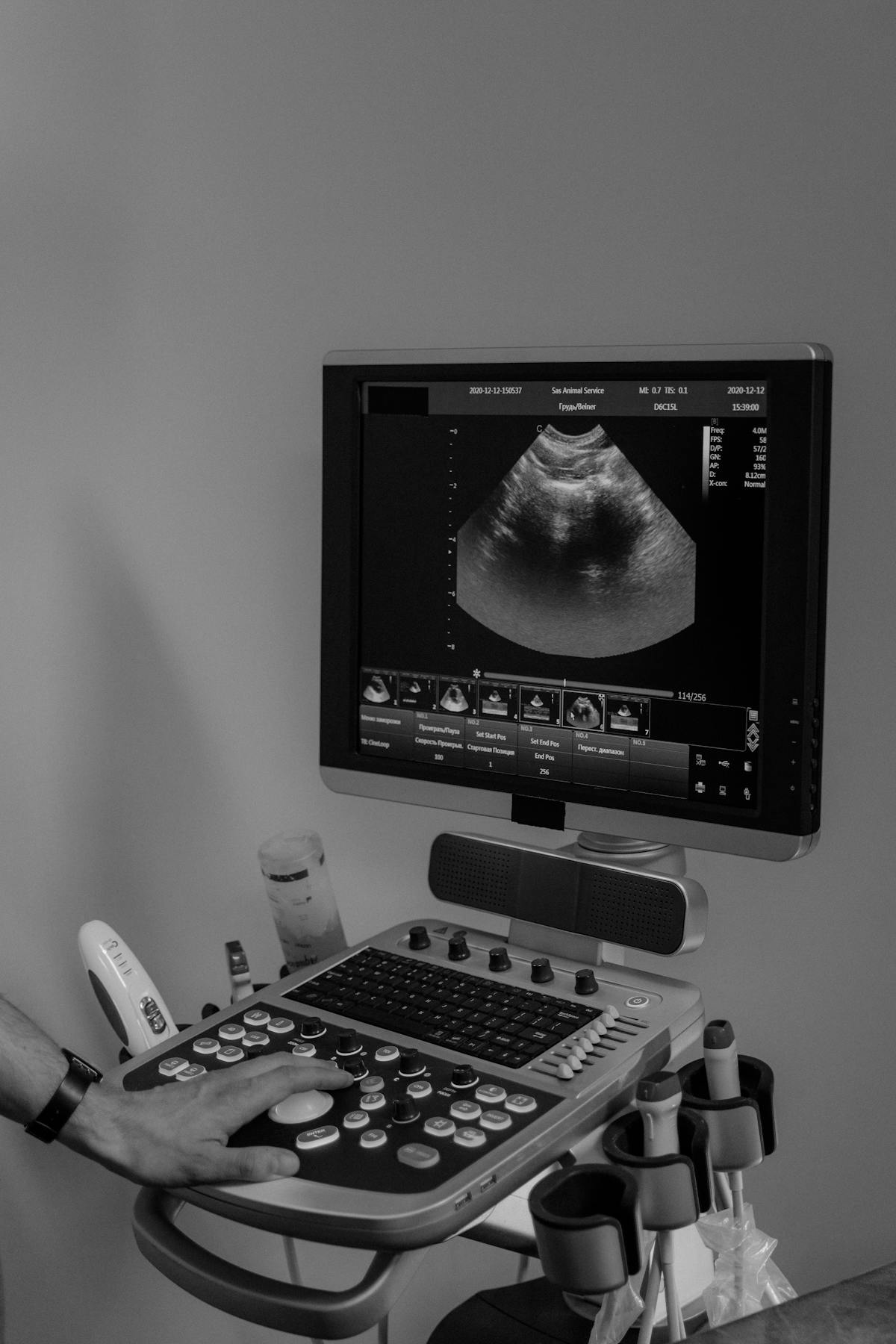
The anatomy ultrasound is a detailed obstetrical examination typically performed at approximately 20 weeks of pregnancy. This scan is an important part of prenatal care and is designed to carefully assess the baby’s growth and developing anatomy.
During this examination, the sonographer obtains a number of required measurements and diagnostic images for radiologist review. Because multiple body systems and structures must be evaluated in detail, this appointment is often longer than other pregnancy ultrasounds.
The anatomy ultrasound may include assessment of:
- Baby’s growth and development
- Brain and facial structures
- Spine
- Heart, lungs and major vessels
- Stomach, kidneys, and other abdominal structures
- Pelvic structures including bladder and genitalia
- Arms, legs, hands, and feet
- Placenta location
- Amniotic fluid levels
- Fetal position and heartbeat
- Maternal cervix and pelvic structures
This examination is highly detailed and depends greatly on factors such as fetal position, movement, gestational age, maternal body habitus, and technical imaging conditions. For this reason, additional images or follow-up imaging may sometimes be required.
Please understand that during the anatomy ultrasound, we may not always be able to show you the fetus or provide viewing opportunities. The primary focus of the examination is obtaining all medically required images and measurements needed for accurate diagnostic interpretation.
For patient comfort and privacy, we allow one guest to join at the end of the examination whenever possible. This allows the sonographer uninterrupted time to focus on the detailed imaging requirements of the scan. Depending on the findings or technical complexity of the examination, guest viewing may unfortunately not always be possible.
During the scan, the ultrasound screen will not be visible while the technologist is performing the examination. This is intentional and allows the sonographer to fully concentrate on capturing the best possible diagnostic images for the radiologist.
Our technologists are highly trained professionals whose role is to obtain the required diagnostic images and measurements. They are unable to provide results or discuss findings during the appointment.
Please note that assessment of the baby’s genitalia (fetal sex) is a routine part of the anatomy examination. If you would like to know the fetal sex (sometimes referred to as the baby’s gender), please inform the technologist at the beginning of your appointment. If the fetal sex can be determined during the examination, the technologist will document this information, and the radiologist can include it in the final report. Technologists are not permitted to provide a written disclosure of the fetal sex at the time of the examination.
Your sonographer will assess and confirm your baby’s heartbeat as part of the examination. Fetal heart rate is typically evaluated using M-mode (Motion Mode) ultrasound, which measures and records the movement of the baby’s heart over time. This allows the heart rate to be accurately calculated while using standard ultrasound imaging.
By comparison, the familiar “heartbeat sound” heard during some scans is produced using Doppler audio, which uses a different ultrasound technique to detect motion and blood flow and convert it into an audible sound. Because Doppler technology uses higher acoustic energy than M-mode, current ultrasound safety recommendations support using M-mode for routine fetal heart rate assessment, especially during the first trimester.
While the heartbeat is carefully monitored and documented to ensure your baby’s well-being, audio playback of the heartbeat is not provided during the examination.
Patients can enjoy free access to both their ultrasound images and reports through PocketHealth following their examination. PocketHealth is a secure online portal that allows you to view, download, and share your imaging records anytime, from any device. This provides an easy and convenient way to keep your pregnancy images and reports accessible throughout your care journey. https://www.pocket.health/en-US/EDI/intro
We understand this ultrasound is often an exciting and emotional milestone for families, and we appreciate your understanding and cooperation as we prioritize accurate imaging, patient care, and the health of both parent and baby.
Obstetric Guest Policy
For the safety, privacy, and quality of your examination, we allow one guest to join at the end of the pregnancy ultrasound whenever possible.
Obstetrical ultrasounds are detailed medical examinations that require the sonographer's full attention and concentration. During the scan, the technologist must obtain many specific diagnostic images and measurements required for radiologist interpretation. Inviting a guest in at the end of the appointment also helps:
- Reduce interruptions during important diagnostic imaging
- Support a calm and quiet examination environment
- Allow the technologist to complete all required measurements efficiently and accurately
Depending on the type of examination, baby's position, or specific findings during the scan, guest viewing may not always be possible.
We understand these appointments are exciting milestones for families, and we appreciate your understanding as we prioritize safe, accurate, and compassionate patient care.
Obstetric Image Access
Patients can enjoy free access to both their ultrasound images and reports following their examination through PocketHealth.
PocketHealth is a secure online portal that allows you to view, download, and share your imaging records anytime, from any device. This provides an easy and convenient way to keep your pregnancy images and reports accessible throughout your care journey.
To access your records, please visit: PocketHealth.
Please note that this service is intended for personal use and secure sharing with your healthcare providers and does not replace the official medical report sent directly to your physician.
Related Pages